Research
How does synaptic plasticity of CA1 pyramidal neurons enable learning and memory in vivo and how does stress influence this process?

We repeatedly inspect the same CA1 basal dendrites using laser-scanning two-photon imaging to detect dendritic spines in mice.
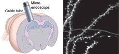
It is widely assumed that synapses are a basic substrate of memory. While two-photon imaging is casting light on the relationship between learning and synaptic turnover in the neocortex, such a relationship is still obscure in the hippocampus. Our lab aims at establishing a direct link between synaptic plasticity and learning in vivo in the hippocampal CA1, and to examine how stress influences this function through some of its established molecular mediators. To this aim, we use optical imaging of dendritic spines in the basal aspect of hippocampal CA1 (Fig. 1) to in vivo investigate long-term synaptic turnover (Fig. 2) learning to perform a memory task that requires the hippocampus.

In vivo time-lapse imaging of a basal dendritic segment of CA1 pyramidal neurons labeled for cytoplasmic GFP expression shows spine turnover during a timespan of 21 days.
Long-term tracking of the same synapses or cells in behaving mice permits for longitudinal studies of the same individuals in different conditions, e.g., prior and after stress or prior and after downregulation/overexpression of specific molecular effectors. This greatly increases statistical power and, importantly, allows direct correlations between molecular, cellular and behavioral phenotypes in the same animal.
How does hippocampal plasticity affect the neural codes underlying representation of experience in CA1?

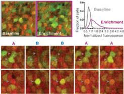
Exploration of a novel enriched environment elicits significant IEG expression. Single sections of in vivo two-photon image stacks show the same CA1 pyramidal neurons after home cage housing or 6 to 8 hours after a 2 hour-long housing in an enriched environment (upper left). Distribution of normalized IEG-driven Venus fluorescence of CA1 pyramidal neurons upon environmental enrichment (magenta curve) is significantly different from baseline (grey curve) (upper right).Exploration of different environments elicits environment-specific activity patterns. Maximum intensity projections of in vivo two-photon image stacks show the same CA1 pyramidal neurons whose fluorescence was induced by exposure to normal environment (magenta arrowheads) or enriched environment (blue arrowheads) (lower).
CA1 representations of the same environment are not stable but turn over with time. We are interested in cellular mechanisms of such turnover and in particular on the influence of regions presynaptic to CA1 on such turnover. To investigate how activity of upstream hippocampal regions affects stability of CA1 representation we probe turnover of CA1 representations repeatedly over long periods of time by in vivo chronic optical imaging by two complementary means: in vivo Immediate Early Gene (IEG) imaging (Fig. 3) or Ca2+ imaging (Ghosh et al., 2011; Ziv et al., 2013) (Movie). We combine long-term imaging of CA1 representations with optogenetic control of CA3 and EC neurons to probe the influence of these presynaptic regions on CA1 representations.


